


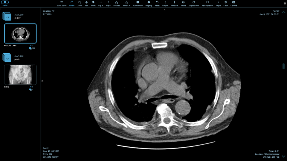
When you open a medical scan on an Online DICOM viewer, you’re not just seeing the image – you’re accessing a treasure trove of hidden medical data. Every DICOM file contains hundreds of metadata fields that tell the complete story behind your scan.
Medical professionals and patients alike can now access this information through web-based viewers, eliminating the need to install specialized software. But what exactly can you see, and how valuable is this data for understanding your medical images?
The Hidden Data Layer in Your Medical Scans
DICOM files work like digital envelopes. The image you see is just the letter inside – the envelope itself contains detailed information about when, where, and how that image was created.
This metadata includes everything from basic patient information to complex technical parameters that radiologists use for diagnosis. Modern online viewers can display most of this information in user-friendly formats.
Think of it as the medical scan’s digital fingerprint. Each piece of metadata serves a specific purpose in the diagnostic process.
Patient Information You Can Access
The most obvious data category involves patient demographics and identification. When you upload a DICOM file to an online viewer, you’ll typically see:
Basic patient details include name, date of birth, patient ID, and gender. Some files also contain weight, height, and pregnancy status when relevant to the imaging procedure.
Medical record numbers and referring physician information appear in most files. This helps ensure the right scan reaches the right doctor and patient.
However, many online viewers allow you to anonymize this data for privacy protection. This feature removes or masks personal identifiers while preserving the medical image and technical parameters.
Study and Series Information
Every medical imaging session generates specific metadata about the examination itself. This category tells you exactly what happened during your scan.
Study dates, times, and unique identifiers help track when the imaging occurred. Series descriptions explain what body part was examined and which imaging protocol was used.
Institution names and department information show where the scan took place. Equipment manufacturer details and model numbers indicate which machine performed the imaging.
| Metadata Category | Common Fields | Clinical Value |
| Patient Demographics | Name, DOB, Gender, Weight | Identity verification, dosing calculations |
| Study Information | Date, Time, Institution, Referring physician | Tracking, workflow management |
| Technical Parameters | kVp, mAs, slice thickness, field of view | Image quality assessment, protocol optimization |
Technical Imaging Parameters
This is where things get interesting for medical professionals. DICOM headers contain detailed technical information about how the image was acquired.
For CT scans, you’ll find parameters like tube voltage (kVp), tube current (mAs), slice thickness, and reconstruction algorithms. These numbers directly affect image quality and radiation dose.
MRI scans include sequence types, repetition times, echo times, and magnetic field strength. These parameters determine image contrast and what tissues appear bright or dark.
X-ray images contain exposure settings, grid information, and detector specifications. Understanding these helps evaluate image quality and diagnostic accuracy.
Measurement and Calibration Data
Online DICOM viewers can extract precise measurement information embedded in the headers. This includes pixel spacing, which tells you the real-world size represented by each pixel in the image.
Calibration data ensures accurate measurements when you use the viewer’s measuring tools. Without this metadata, distance and area calculations would be meaningless.
Some advanced scans include 3D spatial information, orientation markers, and coordinate system references. These enable proper image reconstruction and multiplanar viewing.
Equipment and Acquisition Details
Modern medical imaging equipment embeds extensive technical information in DICOM headers. You can discover the exact scanner model, software version, and imaging protocols used.
Acquisition parameters vary by imaging type but typically include field of view, matrix size, and acquisition time. These affect both image quality and patient comfort during the scan.
For CT scans, radiation dose information appears in the headers. This includes dose-length product (DLP) and CT dose index (CTDI) values, which help track patient radiation exposure.
Contrast and Enhancement Information
When contrast agents are used during imaging, this information gets recorded in the DICOM headers. You’ll find details about contrast type, injection timing, and enhancement phases.
This data is crucial for radiologists who need to understand when the images were captured relative to contrast injection. Different timing phases show different anatomical structures and pathological processes.
Flow rates, injection volumes, and delay times all contribute to proper image interpretation. Without this context, some findings might be misunderstood or missed entirely.
Clinical Context and Procedure Codes
DICOM headers often contain procedure codes, clinical indications, and referring physician notes. This information provides crucial context for image interpretation.
Procedure codes follow standardized formats like CPT or SNOMED, ensuring consistent documentation across healthcare systems. These codes link imaging studies to specific medical conditions or symptoms.
Clinical history sections may include relevant patient symptoms, previous imaging results, or surgical history. This context helps radiologists focus their interpretation on clinically relevant findings.
You can access all this metadata through various Online DICOM viewer platforms, each offering different levels of detail and user interface design.